
Last update images today Decoding The Pelvis: Health Imagery Amp Insights
Decoding the Pelvis: Health, Imagery, & Insights
The pelvis, a bony structure at the base of your spine, plays a crucial role in movement, stability, and overall health. This week, we delve into the "image of the pelvis," exploring its anatomical importance, common imaging techniques, and what those images can reveal about your well-being. We'll also address frequently asked questions to provide a comprehensive understanding. This article is targeted towards anyone interested in understanding their anatomy better, those experiencing pelvic pain or discomfort, and healthcare professionals seeking a refresher.
The Crucial Role of the Image of the Pelvis
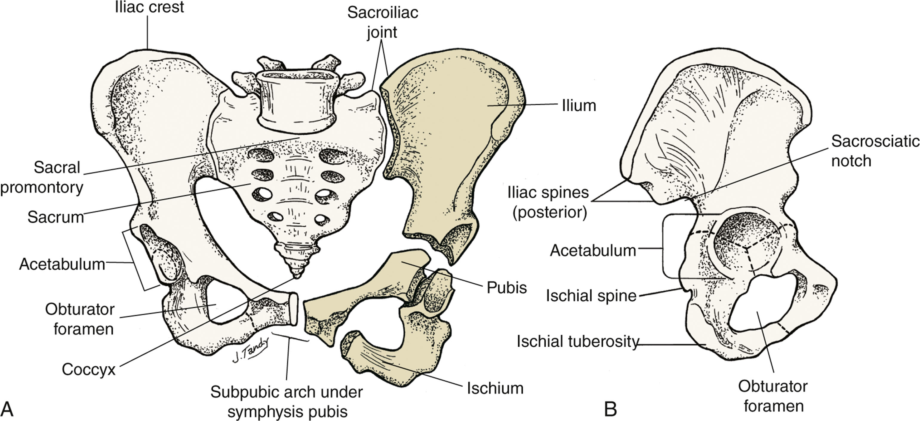
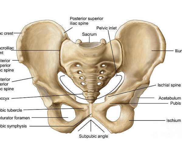
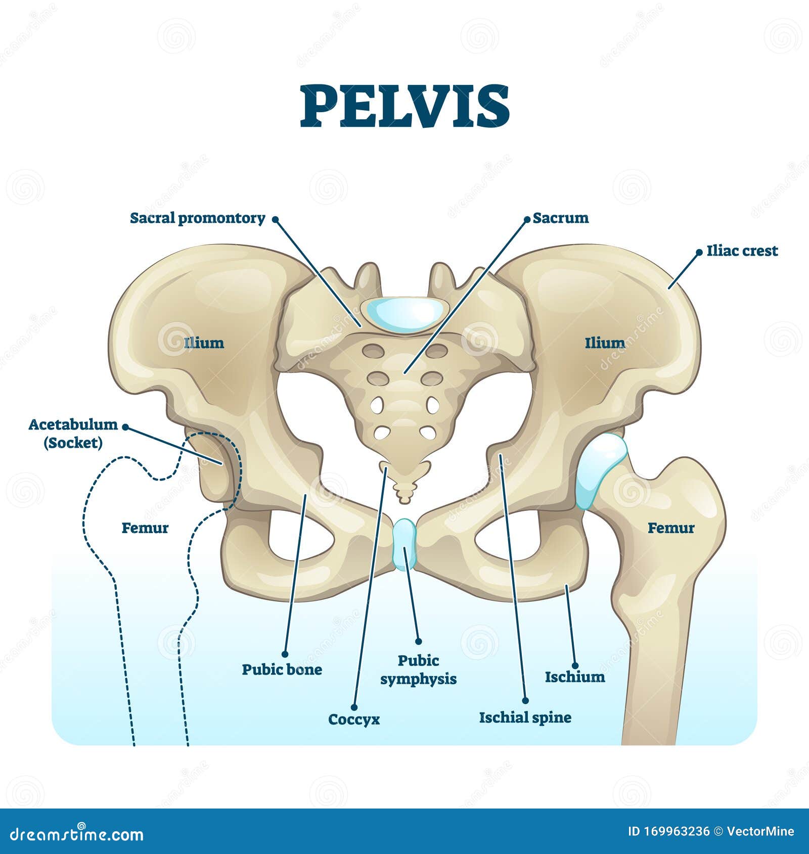
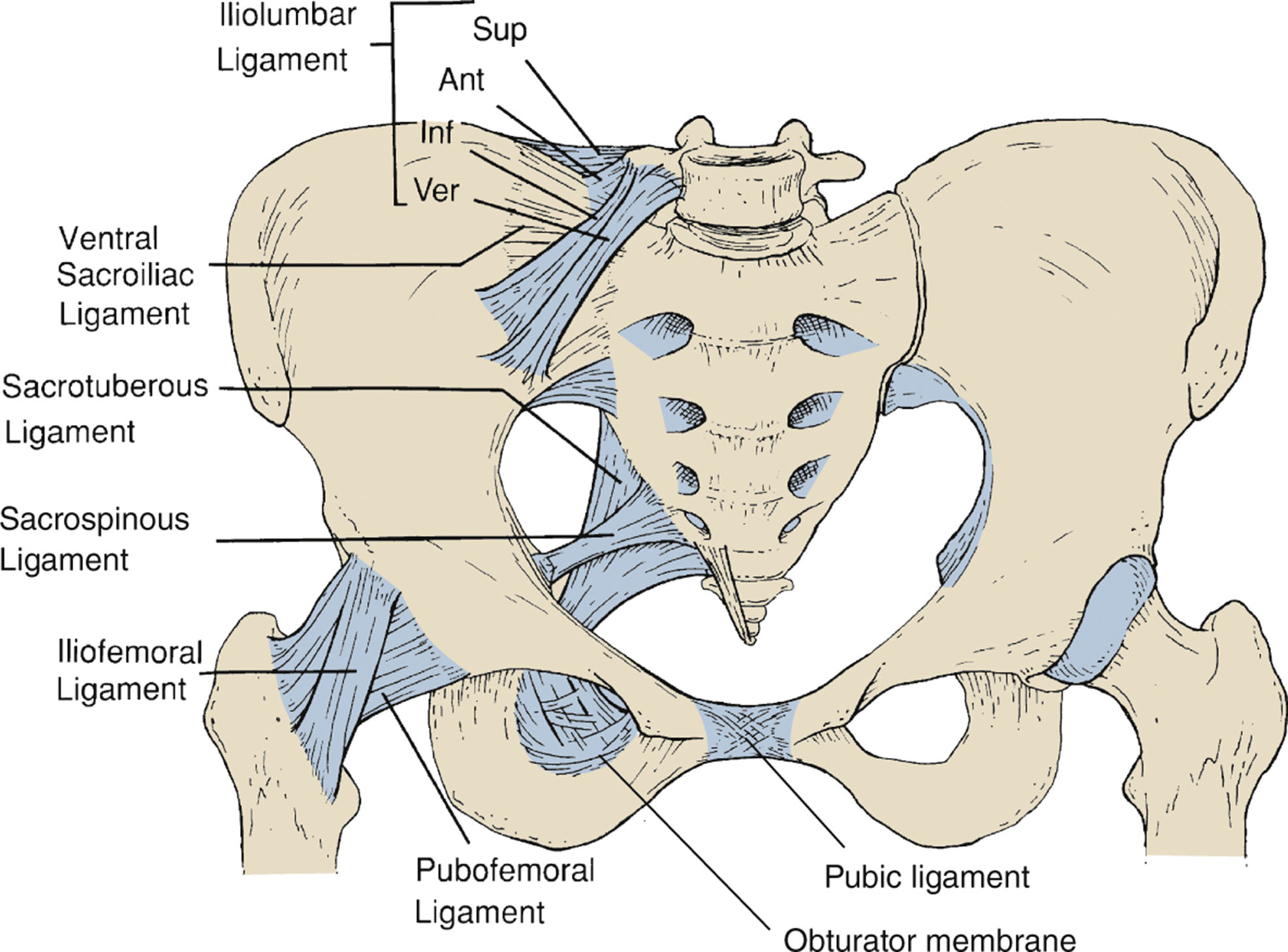
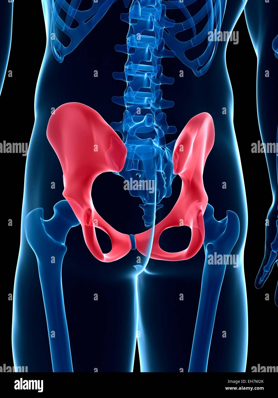
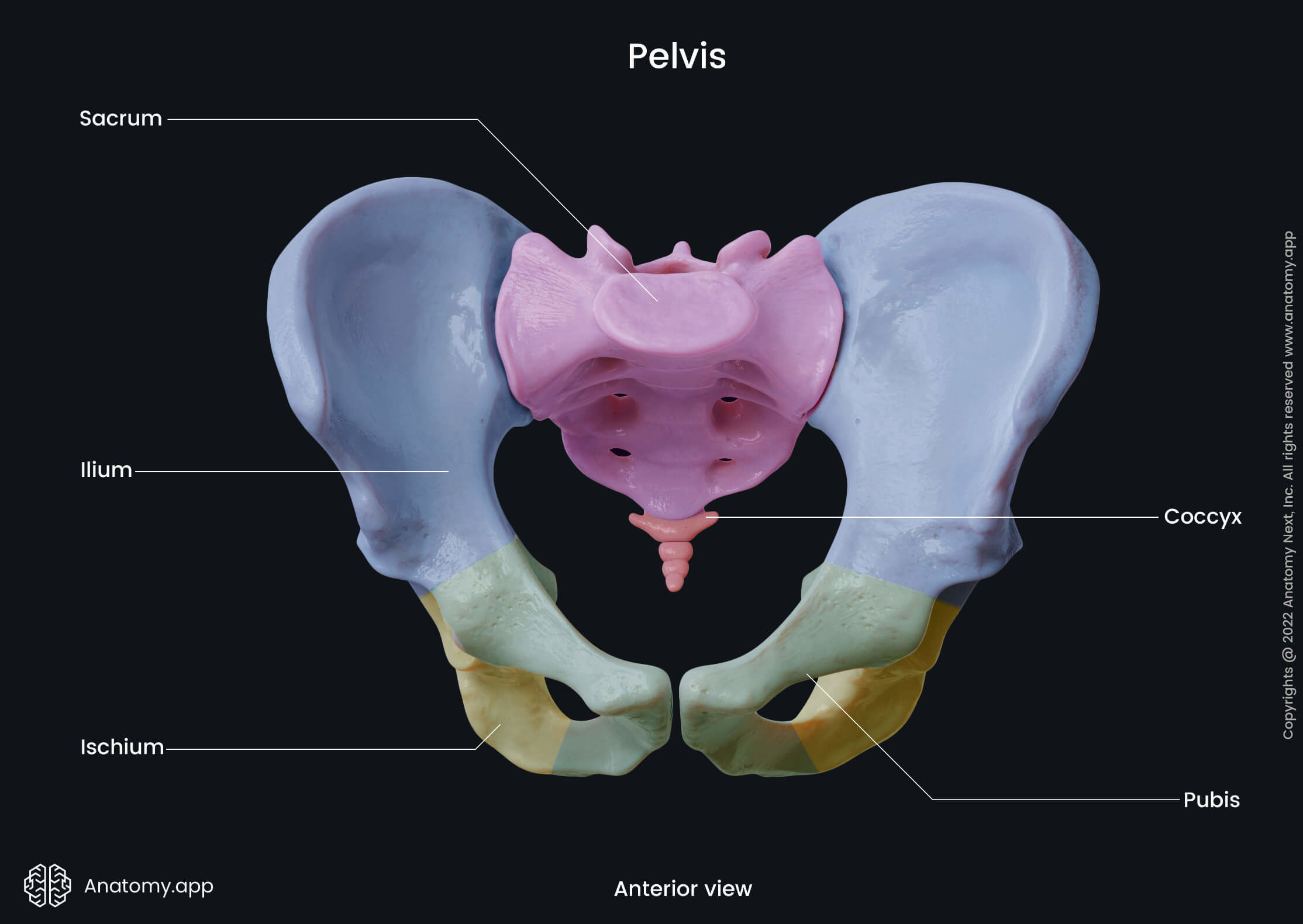
The pelvis is more than just a connection point between your upper and lower body. It houses vital organs like the bladder, reproductive organs (in both males and females), and the lower intestines. Its bony structure protects these organs and provides attachment points for numerous muscles involved in movement, posture, and even childbirth. When we discuss the "image of the pelvis," we're referring to the visual representation of this intricate region, obtained through various diagnostic imaging methods. Understanding the anatomy and function of the pelvis is crucial for interpreting these images accurately.
Why Might You Need an Image of the Pelvis?
Several reasons might prompt a doctor to order an "image of the pelvis." Common reasons include:
- Pelvic Pain: Persistent pain in the lower abdomen, groin, or back.
- Hip Pain: Identifying the source of hip discomfort, which could stem from the pelvis.
- Trauma: After an accident or fall to assess for fractures or dislocations.
- Reproductive Health Concerns: Investigating infertility, abnormal bleeding, or pelvic masses.
- Cancer Screening & Monitoring: Detecting or tracking the progression of certain cancers affecting the pelvic organs.
- Urinary Problems: Exploring potential causes of incontinence, frequent urination, or blood in the urine.
- Inflammatory Conditions: Assessing for inflammatory conditions like sacroiliitis.
Imaging Techniques: Visualizing the Image of the Pelvis
Several imaging modalities can provide a detailed "image of the pelvis." The choice depends on the suspected condition and the level of detail required.
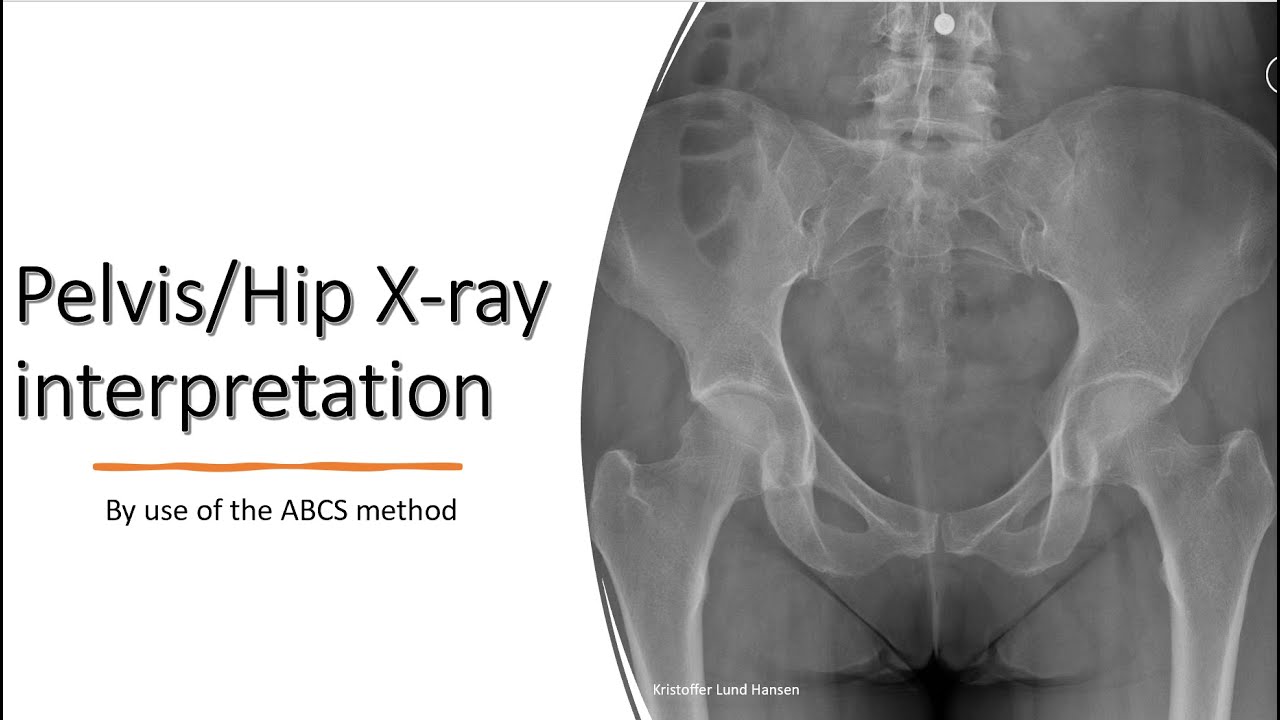
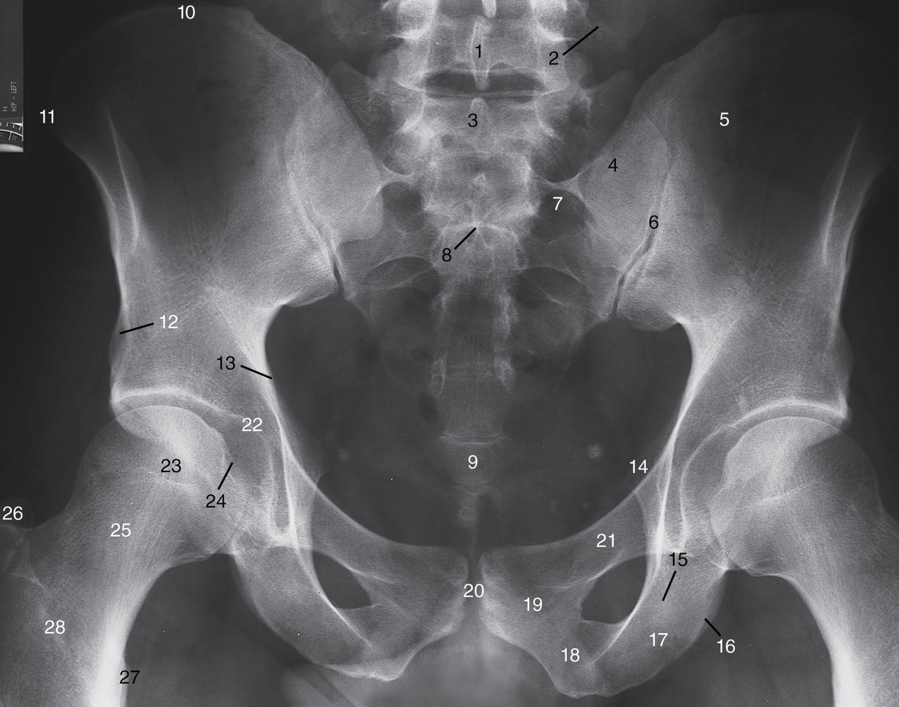
- X-rays: Often the first line of investigation for suspected fractures or dislocations. X-rays are quick, relatively inexpensive, and effective at visualizing bone structures. The "image of the pelvis" obtained via X-ray can quickly reveal breaks or misalignments.
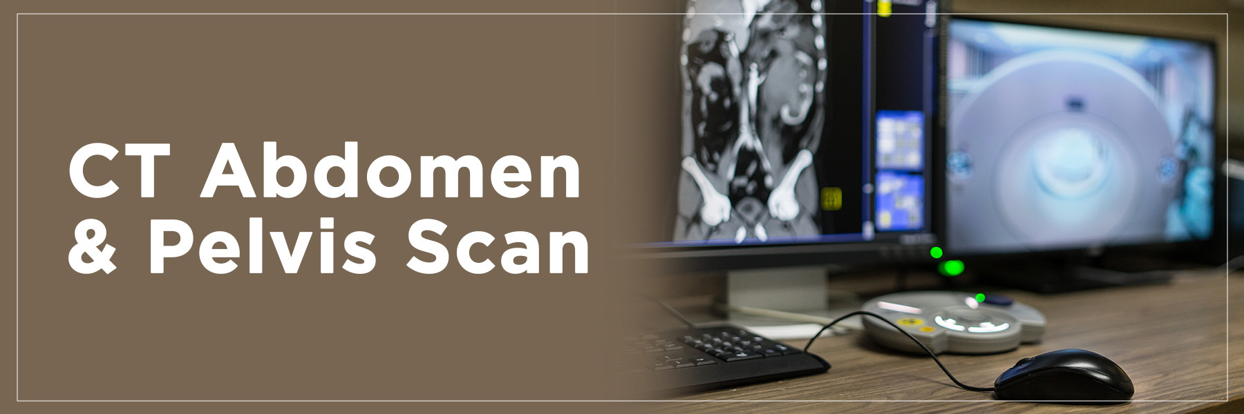
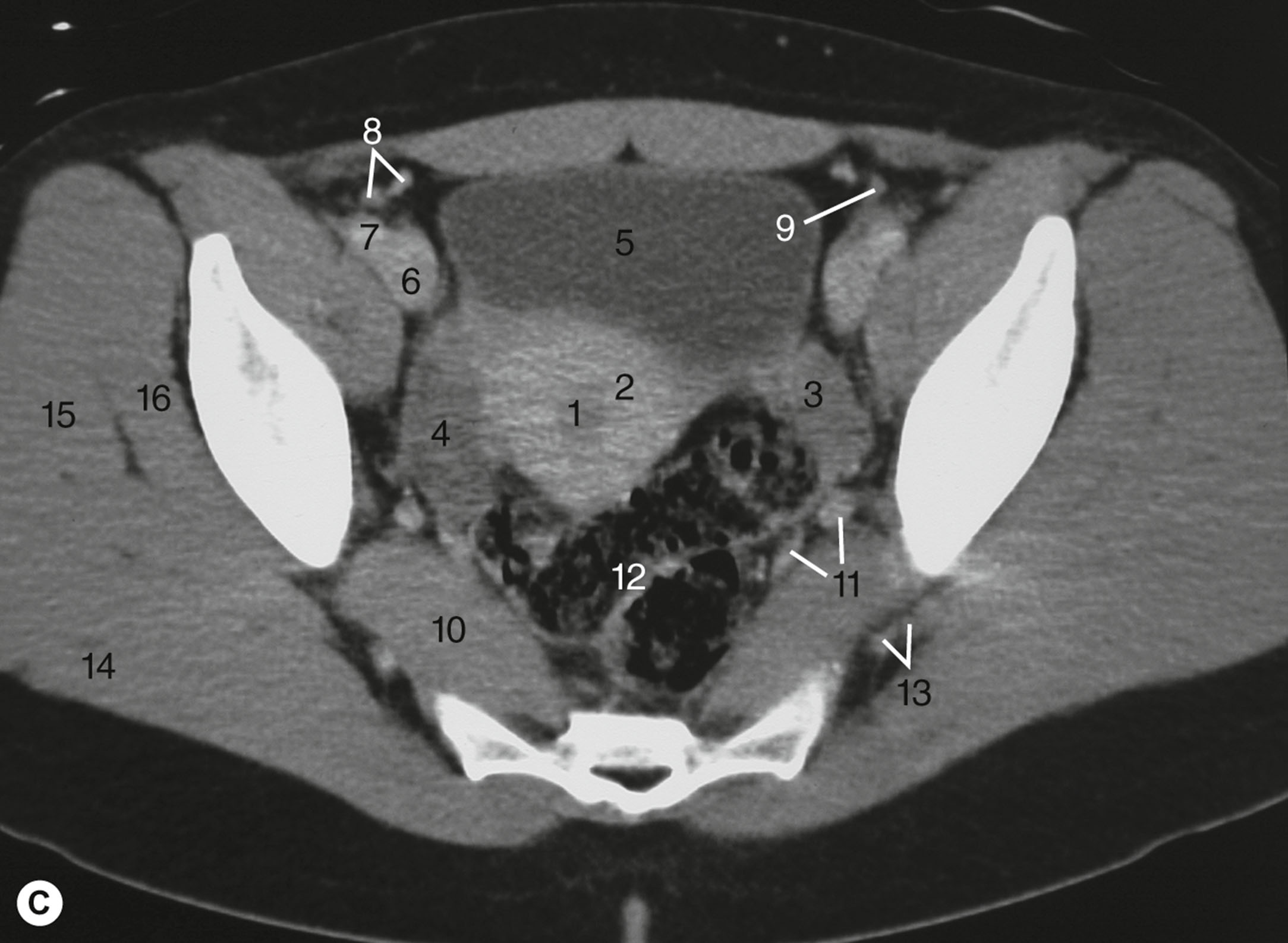
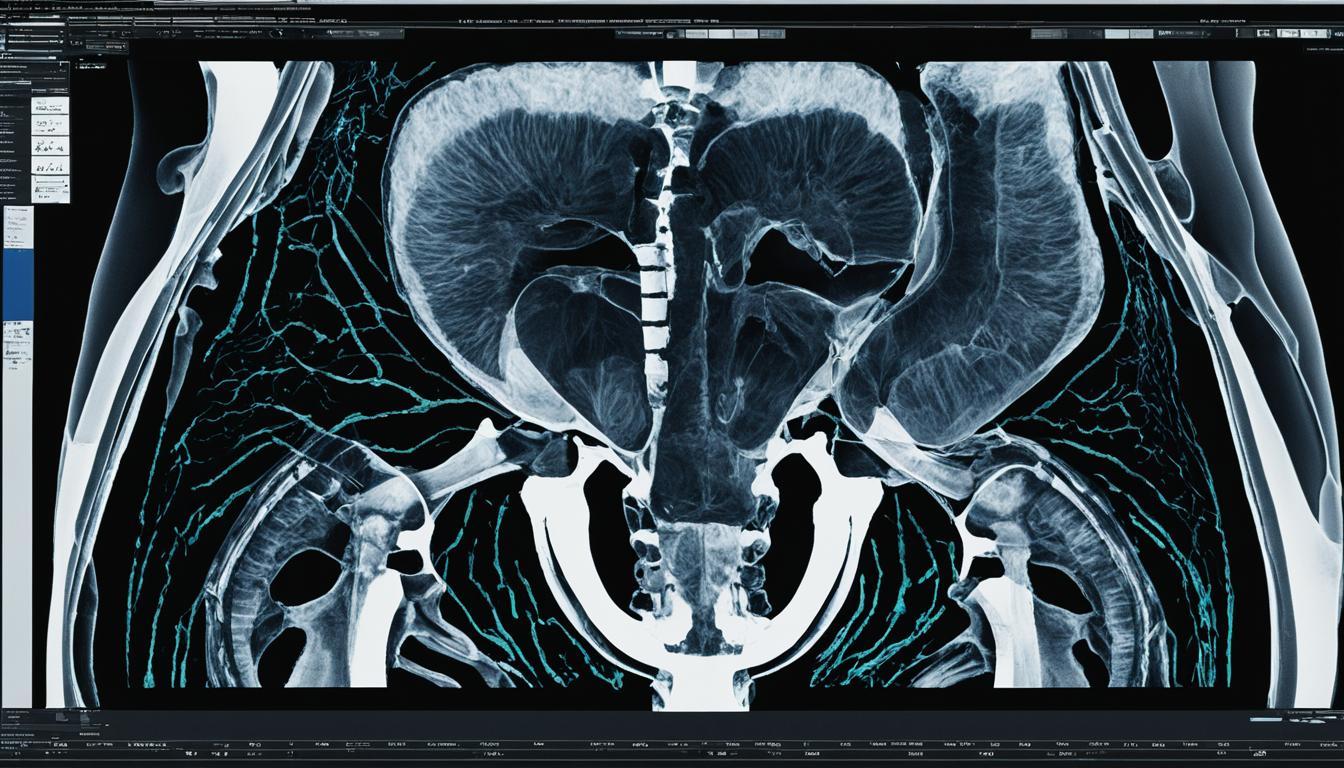
- CT Scans (Computed Tomography): Provide more detailed cross-sectional images of the pelvis, including bone, soft tissues, and blood vessels. CT scans are useful for detecting fractures, infections, tumors, and blood clots. With modern technology, the "image of the pelvis" from a CT scan offers a high level of clarity.
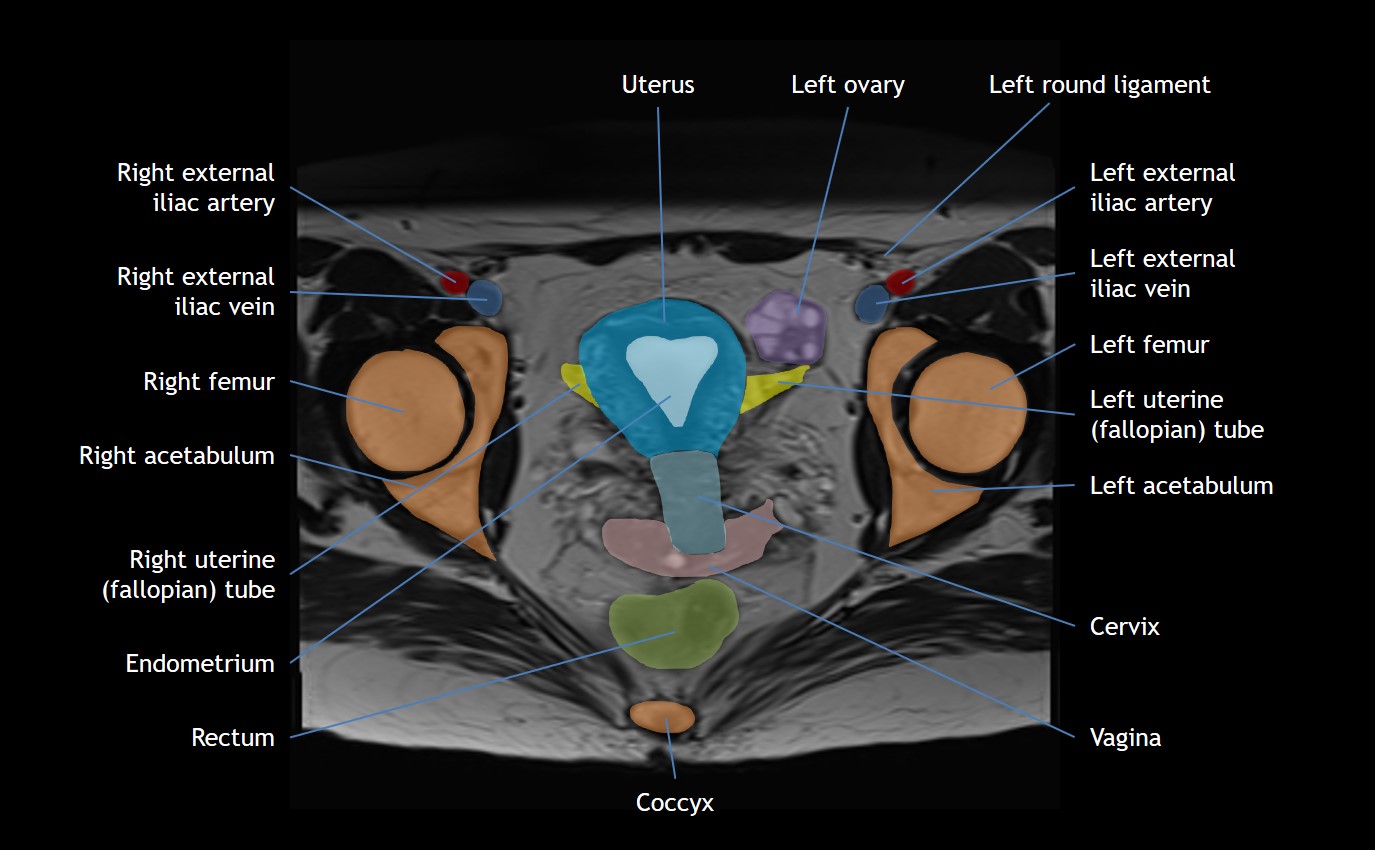
- MRI (Magnetic Resonance Imaging): Uses strong magnetic fields and radio waves to create highly detailed images of soft tissues, including muscles, ligaments, tendons, nerves, and internal organs. MRI is particularly useful for evaluating pelvic pain, soft tissue injuries, and abnormalities of the reproductive organs. An "image of the pelvis" from an MRI provides the best detail for soft tissues.
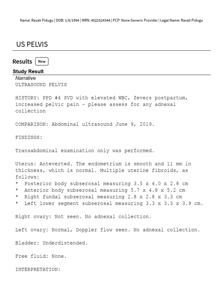
- Ultrasound: Uses sound waves to create real-time images of the pelvic organs. Ultrasound is often used during pregnancy to monitor fetal development and can also be used to evaluate the uterus, ovaries, and bladder. The "image of the pelvis" from an ultrasound is non-invasive and doesn't use radiation.
- Bone Scans: Used to detect areas of increased bone activity, which can indicate fractures, infections, or tumors. A bone scan can provide a functional "image of the pelvis," highlighting areas of abnormal bone metabolism.
Understanding Your Image of the Pelvis: What to Expect
If your doctor has ordered an "image of the pelvis," it's natural to have questions. Here's what you can generally expect:
- Preparation: Depending on the type of imaging, you may need to fast beforehand or drink a special contrast solution. Your doctor will provide specific instructions.
- The Procedure: The procedure itself is usually painless, although you may experience some discomfort lying still for a prolonged period.
- Results: A radiologist will interpret the images and send a report to your doctor. Your doctor will then discuss the results with you and recommend any necessary treatment.
Beyond the Image: Maintaining Pelvic Health
Regardless of whether you need an "image of the pelvis," maintaining good pelvic health is essential. Here are some tips:
- Exercise Regularly: Strengthen your core and pelvic floor muscles with exercises like Pilates, yoga, and Kegels.
- Maintain a Healthy Weight: Excess weight can put extra strain on your pelvis.
- Practice Good Posture: Proper posture helps to align your pelvis and reduce stress on the surrounding structures.
- Listen to Your Body: Pay attention to any pain or discomfort in your pelvic area and seek medical attention if necessary.
- Stay Hydrated: Proper hydration is important for bladder health.
Celebrities and Pelvic Health Discussions
While discussions about pelvic health are becoming less taboo, it's still a topic that isn't often discussed openly. However, some celebrities have used their platforms to raise awareness about pelvic pain and conditions like endometriosis. One example is Lena Dunham, an actress, writer, and producer who has been vocal about her struggles with endometriosis and the hysterectomy she underwent as a result. Her openness has helped to destigmatize these conditions and encourage others to seek help.
Who is Lena Dunham?
Lena Dunham is an American writer, director, actress, and producer best known as the creator, writer, and star of the HBO television series "Girls" (2012-2017). She has been praised for her honest and relatable portrayal of young women navigating the complexities of life. Beyond "Girls," she has written and directed films and authored books. Her openness about her personal health struggles, including endometriosis, has made her a prominent voice in women's health advocacy.
The Future of Image of the Pelvis: Advancements and Innovations
The field of medical imaging is constantly evolving, leading to more advanced and precise methods for visualizing the "image of the pelvis." Some key advancements include:
- 3D Imaging: Provides a more comprehensive view of the pelvic structures, allowing for better surgical planning and diagnosis.
- AI-powered Image Analysis: Artificial intelligence algorithms can assist radiologists in identifying subtle abnormalities in pelvic images, improving accuracy and efficiency.
- Lower-Dose Radiation Techniques: Minimizing radiation exposure during CT scans and X-rays.
Question and Answer about the image of the pelvis
-
Q: What is the image of the pelvis used for? A: To diagnose and monitor a wide range of conditions affecting the bones, joints, muscles, and organs within the pelvic region.
-
Q: What are the common imaging techniques used to create an image of the pelvis? A: X-rays, CT scans, MRI, ultrasound, and bone scans.
-
Q: What should I expect during a pelvic imaging procedure? A: Expect to lie still during the procedure, and you may need to follow specific preparation instructions, such as fasting or drinking a contrast solution.
-
Q: How can I maintain good pelvic health? A: Exercise regularly, maintain a healthy weight, practice good posture, and listen to your body.
Summary: The "image of the pelvis" is crucial for diagnosing and monitoring various conditions. Imaging techniques like X-rays, CT scans, MRI, and ultrasound provide detailed visualizations of the pelvic region. Maintaining pelvic health through exercise and a healthy lifestyle is also essential.
Keywords: image of the pelvis, pelvic pain, pelvic floor, MRI, CT scan, X-ray, ultrasound, pelvic health, bone scan, Lena Dunham, endometriosis, pelvic anatomy, pelvic imaging.


Pelvic Anatomy Bone Ligaments OrthoFixar 2025 Pelvic Anatomy 1 Pelvis Anatomy High Res Stock Images Shutterstock In 2025 Pelvis 22c814a11c7835d5cccd7adc1121f707 Leiden Drawing Pelvic Inlet And Outlet English Labels AnatomyTOOL Lumc Data Item Download FileUs Pelvis 2 Jan 2025 PDF 1Anatomy Bony Pelvis And Lower Limb Pelvic Bones Treatment 11323Ct Abdomen And Pelvis With Contrast Cpt Code 2025 Phil Gray Cpt Code Ct Abdomen And Pelvis With Contrast 5 Facts About The Anatomy Of The Pelvic Cavity Pelvic Cavity Girdle Anterior #keepProtocol
The Lumbo Pelvic Hip Complex 2025 LPHC Breakdown Master The Lumbo Pelvic Hip Complex Pelvic Girdle Diagram Labeled F16a3bd9 93f3 43cb B8dc 9c59d0eefc37 Large Pelvis Definition Anatomy Functions Lesson Study Com 7.192 112666 CT Abdomen Pelvis Scan Purpose Preparation Diagnosis CT Abdomen Pelvis Scan Mri Female Pelvis Anatomy Axial Image 4 Pelvis Anatomy Pelvi U010 028b 9781437736540 Human Pelvis Illustration Stock Photo Alamy Human Pelvis Illustration EH7M2K Pelvis Anatomical Skeleton Structure Anatomy Of The Pelvis Superior Pelvis Anatomical Skeleton Structure Anatomy Of The Pelvis Superior View And Inferior View Medical Education Scheme 2WAXY7W Diagram Of Pelvic Area Anatomy Of Human Pelvic Bone Stocktrek Images
Pelvis Photograph By Medical Graphics Michael Hoffmann Science Photo 3 Pelvis Medical Graphicsmichael Hoffmannscience Photo Library The Pelvis Radiology Key F06 01 9780443105609 2025 New Female Pelvis Model Life Size Female Pelvic Skeleton 41RA RLVSWL The Pelvis Radiology Key F06 03 9780443105609 The Pelvis Radiology Key F06 02 9780443105609 Pelvis Anatomy Xray Hq720 Pelvis Photograph By Medical Graphics Michael Hoffmann Science Photo Pelvis Medical Graphicsmichael Hoffmannscience Photo Library DIAGRAM Femur And Pelvic Bones Diagram MYDIAGRAM ONLINE Pelvis Anatomical Skeleton Structure Labeled Vector Illustration Diagram Medical Education Scheme Ilium Ischium Coccyx Sacrum 169963236
Pelvic Injuries RCEMLearning Pelvis Scaled Levator Ani Muscle Group Location And Lesser Pelvis Bones Outline 406aa31ab34bb70ecd473f7528e2e9cc PDF Imaging Of The Abdomen And Pelvis 2025 03 23 Carelon Clinical PDF Imaging Of The Abdomen And Pelvis 2025 03 23 Pdf 86975 1080x675 3D Illustration Of Pelvis Medical Concept Stock Illustration D Illustration Pelvis Medical Concept Part Human Skeleton 71822802 The Pelvis Radiology Key F06 36c 9780443105609 OTs In Pelvic Health Summit 2025 53aeca 5eb1 Cfd 43a6 4ee5a7b3f5da Social Media Ads 2024 Updated Idea 10
Pelvis Reconstruction Plate Market 2025 Market Shaping Insights With 1 1743667224649Pelvis Anatomy Module CaseStacks Com Fpelaxw10 Pelvis Anatomy Color Structure Pelvic Skeleton Vector Image Pelvis Anatomy Color Structure Pelvic Skeleton Vector 31602401